

Posterior Leg Muscle Anatomy
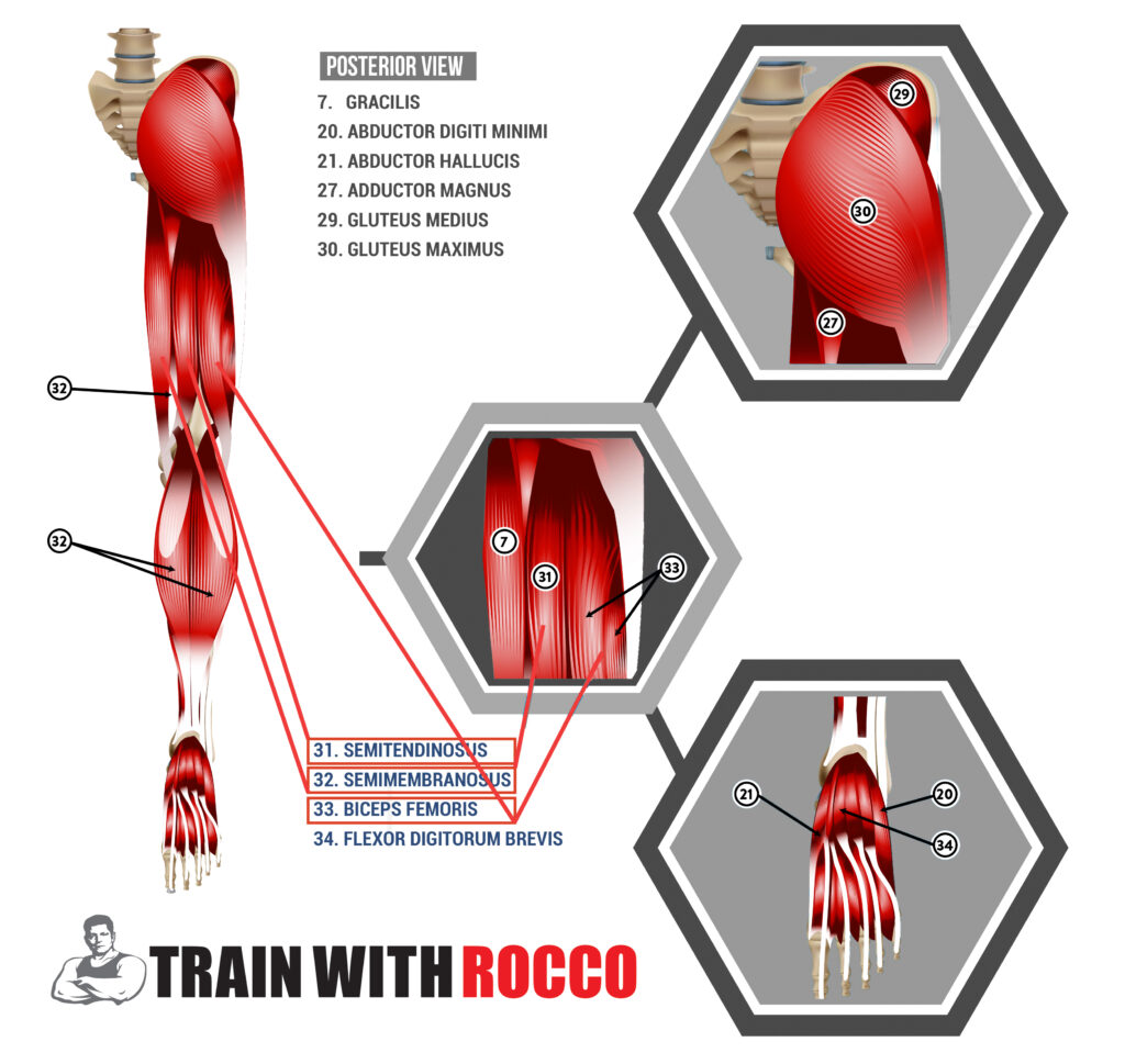
Your leg muscle anatomy begins with the glutes, which make up the buttocks. The glutes are the gluteus maximus, medius, and minimus, and a group of muscles known as the six outward rotators. The piriformis, an essential muscle when it comes to sciatic pain, is the only muscle from the outward rotators that you need to be aware of. Collectively referred to as the glutes or the abductors.
Adductors
The adductors on the inner thighs serve as the glutes’ stabilizing muscles. Long and short muscles make up the adductors. The thin gracilis inside the thigh and the adductor magnus, which extends to the knee, are two examples of lengthy adductors. The pectineus, adductor brevis, and adductor longus are the short adductors. The iliacus and iliopsoas, two muscles located deeper in the pelvis, also assist with adduction.
Hip Extensors
Let’s now examine the hip extensors. The Glute max drives the leg behind you because it is a hip extensor. In one type of hip extension, the hip is extended back behind you by the deeper-lying hamstring muscles on the back of the thigh. Hip extension also occurs when your leg is extended in front of you, pulling it back down with a straight leg. The hamstring’s top fibers are active in such movement.
Hip Flexors
The deep hip flexors serve as counterbalancing muscles for the hip extensors or higher hamstring fibers. Flexion pulls the leg upward and forwards, while extension puts the leg behind. The primary hip flexors are the human body’s only muscles extending from the spine to the upper leg. Many people carry a lot of tension in those muscles, which will cause them to exert a lot of torque on the lower back. The psoas and the iliopsoas, connected to the iliacus muscle, are the most significant muscles you’ll learn to isolate with Resistance Stretching.
Most people do not distinguish between the upper and lower hamstring fibers, I do because we have different exercises for each. Knee flexion, often known as bending your knee, involves your lower hamstrings.
Knee flexion also benefits from the calf muscle called the gastrocnemius. The calf is referred to as the secondary knee flexor. It has a lot of muscle that attaches to the femur and runs behind the knee. Although you may not want it to flex your knees, you can’t ignore it because it is coordinated enough to perform some of that motion.
Anterior Leg Muscle Anatomy
The Quadriceps
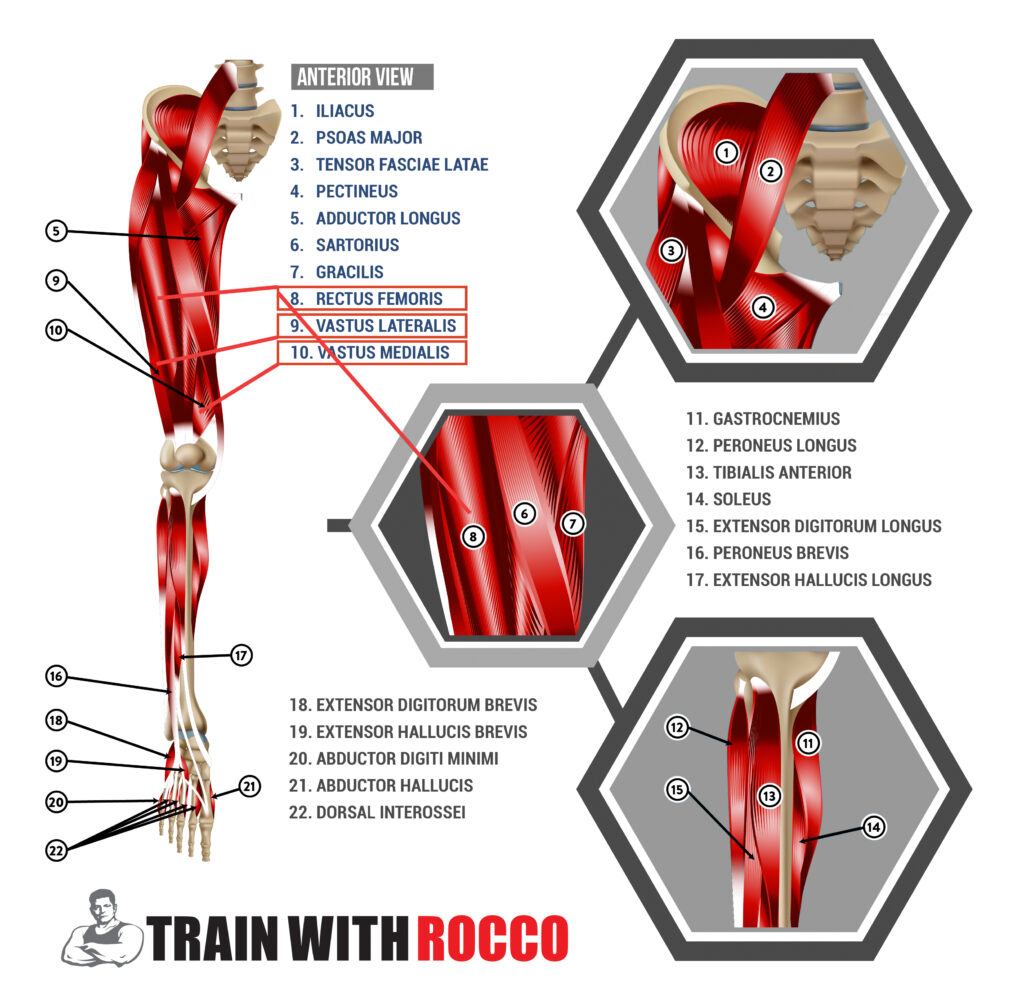
The balance muscle group for knee flexion is the four muscles that make up the front and bulk of the thigh quadriceps. They are located in the middle to lower part of the leg. The rectus femoris, vastus intermedius, vastus medialis, and vastus lateralis are among them. Only one muscle, the rectus femoris, can be utilized to flex the hip slightly: it crosses the hip joint. We refer to it as a hip pointer when you remove or rip that attachment off the front of the pelvis. Many people will overrecruit the rectus femoris and the tensor fasciae latae (TFL) and overextend the psoas, which can lead to injury.
TFL vs IT Band
Many people mistakenly believe they are rolling out their IT band when using a foam roller. The actual bothersome muscle is your vastus lateralis, which is located beneath that little tendon and is referred to as the IT band. It’s a sign that the TFL, the gluteus medius, and the tissue on the outside of the hip are tight if you experience issues like IT friction syndrome, which produces a clicking sensation at the IT band across a portion the knee. It’s as if these tissues are the instrument’s top screws and have overtightened the IT band or the guitar string.
Too much tension in the TFL and gluteus medius causes the IT band to experience rotational issues at the knee. Which can cause undue wear and tear. However, having a tight IT band does not necessarily make rolling on your thigh’s side more painful. It helps to have tight quads, especially the vastus lateralis.
Plantar Flexors & Dorsiflexors
Your plantar flexors are the muscles that allow you to point your foot. Which are gastrocnemius and soleus muscles in your lower leg. Those are the primary plantar flexors. When you walk, they serve as your propulsion muscles. Other tissues, like the popliteus, a stabilizing muscle behind the knee, and the plantaris tendon, which extends to the Achilles, also participate in this function.
Plantar flexors point your foot, and dorsiflexors flex it. The dorsiflexors are located on the opposing side. These include the longest extensor digitorum and anterior tibia.
Functional Planes
Understanding the functional planes is beneficial because movement involves different planes. As a result, it is wise to consider dividing the body in half, both vertically and horizontally. The sagittal plane is where all forward motion takes place. That might include both extension and flexion. Sideway movements occur in the coronal plane as well as flexion and extension Rotation uses the transverse plane.
You can decide which muscle line you want to stimulate. So you can exercise in a way that compels those muscles to work along that vector.